Sunday 26 June 2022

Summary


Leptospirosis is one of the important diseases in Malaysia. The study of leptospirosis is extensive and has also been studied in various countries. This portfolio comprises the history and impact of Leptospirosis on Malaysia’s economic and social value. The transmission and symptoms levels are also discussed as well as the possible cure and methods of identifying the disease. Lastly, prevention steps prepared according to the guidelines of the Ministry of Health Malaysia and policies are also included in this portfolio. 


Click here if you want to read in magazine form


Friday 24 June 2022

1.1 Definition of Leptospirosis

1.1 Definition of Leptospirosis

Leptospirosis is an example of zoonotic disease (spread from animals to humans) for which globalization and social variation have different epidemiological patterns for the poor and rich community. According to the Malaysia Ministry of Health (KKM) official website, the other names for this disease are Weil’s disease, Icterohemorrhagic fever, Swineherd’s disease, Rice-field fever, Cane-cutter fever, Swamp fever, and others. This disease is a significant health problem in developing countries especially for the poor rural society (Bharti, 2003). It has been reported that this disease commonly occurs in tropical and subtropical countries that are suitable for the bacteria's growth and it can reach more than 1 million cases per year globally. Malaysia is one of the Southeast Asia countries that has a significant increase in disease outbreaks and reported cases in recent years (Garba, Bahaman, Khairani-Bejo, Zakaria, & Mutalib, 2017). Until now, Malaysia is endemic for Leptospirosis with increasing cases recorded over the years.

Leptospira Bacteria is a photograph by Kateryna Kon/science Photo Library which was uploaded on October 1st, 2018.

Electronic image of Leptospira interorgans


Leptospirosis disease is caused by the spirochetes bacteria belonging to the genus Leptospira. These bacteria are gram-negative and aerobic bacteria. The common size of bacteria is 20 micrometers long and it has a right-handed helical (Karpagam & Ganesh, 2020). Sixty-six different species of Leptospira have been discovered and it has been divided into 2 clades and 4 subclades (P1, P2, S1, S2) according to their genomic sequence. 8 of 19 species from the P1 (L. borgpetersenii, L. alexanderi, L. interrogans, L. mayottensis, L. kirschneri, L. noguchii, L. weilii, & L. santarosai) can cause severe disease and 21 species from P2 were believed could cause mild disease in human. Other 26 species belong to the S1 and S2. They are saprophytes that undergo saprotrophic nutrition (Caimi & Ruybal, 2020). Rodents, cattle, dogs, horses, sheep, swine and wild animals are believed to be the reservoir hosts of the bacteria (Ellis, 2015).

1.2 History of Leptospirosis in Malaysia


        In April 1925, Fletcher encountered by chance the first case of leptospirosis in the Institute of Medical Research, Kuala Lumpur, Malaysia 10 years after the isolation of Leptospira for the first time from human patient blood in Japan (Inada, Ido, Hoki, Kaneko, & Ito, 1916). Fletcher also identified 3 different serovars (Leptospira interrogans serovar Icterohaemorrhagiae, Hebdomadis, and Pyrogenes) after isolating the pathogen from the liver, blood and kidney from the 21 patients in Malaysia. The blood from a patient with unknown origin fever was inoculated into guinea pigs and it developed jaundice and hemorrhages in the nose. The guinea pig died 13 days after inoculation and the postmortem result showed signs of leptospirosis. Fletcher also introduced a new medium in semi-solid form that is widely used in many laboratories nowadays for the isolation and maintenance of the leptospira culture (Fletcher, 1928).


Picture of two cultures of Leptospira sp. strain Manara. Two cultures of Leptospira sp. strain Manara in Fletcher medium supplemented with 3% (left tube) and 1% (right tube) seawater after 48 h of incubation at 28°C. The dinger rings corresponding to Leptospira sp. growth is indicated with red arrows. doi:10.1371/journal.pone.0144974.g001

In the earlier stage of work regarding the Leptospirosis in Malaysia, it only focused on the patients who were in the military hospital. In 1957, it was reported that 614 military personnel and 238 civilian patients admitted at the military hospital were suffering from the fever. Based on the serology test, 34.7% and 13% respectively were confirmed to have Leptospirosis (McCrumb et al, 1957). There was also a previous study that concluded that rubber plantation laborers and the workers at sewage, drainage, forestry, and town cleaning as well as antimalarial workers were exposed to high risk of Leptospirosis infection and become the an occupational disease (Shafie et al, 2012). However, travel to endemic areas and recreational activities such as water sports have increasingly been reported as a major risk for this infection (Victoriano et al, 2009). 

In 2014, it was reported that the peak of the leptospirosis cases was 7806 confirmed cases with 92 deaths (Zainudin, 2015). It does not include the cases that may be under-reported or misdiagnosed and it can be considered as a significant public health threat locally. In 2016, it has been reported that forty-two patients with confirmed diagnosis of leptospirosis in Perak. The case fatality rate was 14.3%. Malay ethenic was the most affected by the diseases and majority of them were male (Fann, Vidya, Chong, Indralingam, & Christoper Chan, 2020). Minly, the outbreak would occur after the effect of heavy rainfall or flood season and it can be a seasonal incidence.


Thursday 23 June 2022

1.3 Transmission of the disease


Leptospirosis is transmitted by pathogenic or saprophytic Leptospires (World Health Organization, 2003). Pathogenic and saprophytic leptospires can both be found in the renal tubules of animals that are hosts, while saprophytic leptospires are more common in damp or tropical climates. Saprophytic leptospires species contaminate water ways without necessitating a host, thrive only on organic matter, and do not infect humans (The Leptospirosis Information Center, 2010). Pathogenic Leptospires, on the other hand, require a host to reproduce (Institute for International Cooperation in Animal Biologics, Center for Food Security and Public Health. Leptospirosis, 2005). Pathogens can be transmitted into the ecology through the urine of animals carrying pathogenic Leptospirosis. The rodent hosts that contain this pathogenic leptospire suffer no harm. They are often described to as "natural hosts."

In a polluted environment, leptospirosis can be spread directly or indirectly from one host to another. When an animal's excrement is expelled, it pollutes the soil and water. After consuming contaminated water, humans are impacted. The dangerous Leptospires bacterium can potentially be passed on to people through contaminated food. By coming into direct touch with Leptospires through wound infections, food, and drink, the individual can be infected. When exposed to dirty water for an extended period of time, it can enter the human body through the nasal, oral, and ocular mucosa. After penetrating the In the 2000 Eco-Challenge multisport race in Malaysian Borneo, a large number of competitors experienced leptospirosis. The Centers for Disease Control and Prevention interviewed 189 people, and 80 (42%) of them fulfilled the leptospirosis clinical diagnosis. Exposure to the rain-swollen Segama river for long periods of time was one of the risk factors (photograph (a) credit Reed Hoffmann). Whereas in B section, its portrayed there was a small community in an area is a typical leptospirosis epidemiologic environment. Citizens of tropical regions with significant rainfall are more likely to contract leptospirosis, especially if residual water is contaminated with excrement from wild or domesticated animals, which can operate as intermediate hosts for pathogenic Leptospira species.

Leptospirosis can also disseminate through the airways. When Leptospires in urine dissipate into air droplets and are inhaled by individuals, they can lead to disease. Even though human-to-human disease transmission is unusual, it should be recognised that consensual sex and nursing could allow pathogenic Leptospires to passed from one generation to another (Lim, 2010). In addition, an infected woman's unborn child may be exposed to hazardous Leptospires. Individuals can expel leptospires in their urine for up to eleven months. Rodents and wild animals such as cattle, pigs, rats, buffaloes, and dogs, which develop leptospires in their urinary for months or years, are a reservoir of pathogenic Leptospires. The predominant carriers of pathogenic Leptospires would be all these animals.

Leptospira may persist in the environment for long periods of time because of the significant dampness and moderate temperatures. In particular, strong rains and numerous floods, especially during the monsoon season, intensify the leptospirosis outbreak. Leptospirosis, on the other hand, has a seasonal distribution, with the bulk of reported cases during the rainy season and following floods (Benacer, 2016). The majority of cases are caused by occupational exposures and recreational activities. Veterinarians, zookeepers, pet store owners, butchers, and other professionals who work with wild animals on a daily basis are more vulnerable. Military personnel, farmers, fishers, and plumbers, for instance, engage in situations that include water, mud, and sludge, and are thus particularly vulnerable human body, leptospires invade the circulatory system and attack bodily tissues and organs.


1.4 Stage level and symptoms of the disease


Leptospirosis is divided into two categories. First category is know as mild leptospirosis that is the most common type of leptospirosis, accounting for 90% of cases. Muscle soreness, chills, and even a headache are some of this category symptoms. Second is extensive leptospirosis which can develop in between 5% and 15% of cases. If the bacterium affects the liver, kidneys, or other vital structures, it can cause organ damage, internal haemorrhaging, and fatality.

Leptospirosis usually takes 5-14 days to develop, although it can span anything else from 2 to 30 days. In contrast, patient, does not display clinical symptoms in less than 24 hours except if the volume of germs penetrating the body is bigger than normal. In most cases, the infection is systemic and affects the entire body. Anicteric leptospirosis and icteric leptospirosis are the two forms of infection that present with diverse clinical manifestations (Madhu & Aparna, 2008)

Anicteric leptospirosis is a type of leptospirosis that does not induce jaundice in 90% of cases (Lim et al., n.d.). Anicteric leptospirosis patients go through phases 1 and 2 of the illness. The sickness is invariably associated with aseptic meningitis in the anicteric subtype of leptospirosis (Madhu & Aparna, 2008). It a little less severe form of leptospirosis is rarely lethal, but it can trigger pulmonary bleeding and mortality even if there is no jaundice. Flu-like characteristics such as severe headaches, rapid fevers of 39°C or higher, eyes inflammatory, muscle aches, diarrhoea, lethargy, nausea and vomiting, chills, rigours, and maculopapular rashes are present in phase 1, also known as acute or septicemic phase.

Anti-leptospira antibodies proliferate during phase 2 (the immunological phase), allowing leptospirosis pathogens to be discovered in the patient's urine output. Infected individuals become ill again during this phase, which can last up to 30 days or longer (Leptospirosis Information, 2021). Some people, however, do not progress to the second stage of the illness. The remaining 10% of patients progress to icteric leptospirosis, often known as Weil's syndrome (Lim et al., n.d.). The second phase of anicteric leptospirosis is far more severe than the first. The bulk of the clinical manifestations are identical to anicteric leptospirosis, however the sickness can progress to be lethal. Within 10 days, organs such as the liver, kidneys, brain, heart, and central nervous system are affected.


Leptospirosis in its clinical phase was observed in figure above. At section A, a 37-year-old man who raised pet rats developed subconjunctival haemorrhages and icterus, as well as fever, myalgia, and severe headache. He had impaired liver and kidney functionality when he was hospitalized. One week after manifestation, serological tests for leptospiral antibodies shifted from negative to positive. He was given injectable penicillin and made a full recovery (Haake & Levett, 2014). Whereas at setion B, its reported that A 50-year-old guy experienced a devastating lung haemorrhage after returning from a trip in Malaysia, where he had wandered through mangrove woods. On the second day of admission, the patient's respiratory condition deteriorated, necessitating mechanical respiration and a blood transfusion. He was given doxycycline first, then amoxicillin, and he recovered slowly yet entirely. Four months following injection

2.0 Strategies


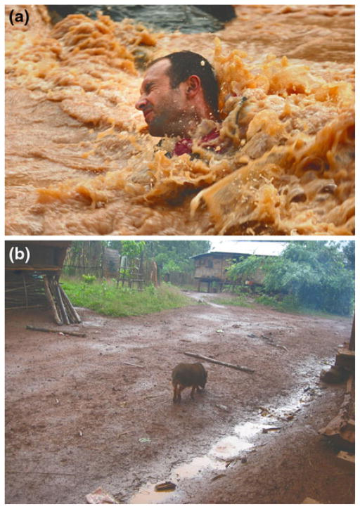
 2.1 Identification of the disease

After the formation of leptospira-specific antibodies, initially primarily of the IgM class, leptospira are present in the blood until they are eliminated after 4–7 days. IgM antibodies can persist for many months, which raises the issue of whether a positive IgM result truly detects a present infection. This is a drawback of IgM detection assays. There are five techniques used to identify Leptospira disease which are rapid leptocheck test, IgM ELISA test, microscopic agglutination test (MAT), real-time PCR assay, and blood culture. 


2.1.1 Rapid leptocheck tests 

Following the idea of immunochromatography, a special membrane-based two-site immunoassay. The anti-human IgM colloidal gold conjugate combines with the IgM antibodies in the sample set as it passes through to the membrane of the testing machine. This compound is bound by the leptospira genus particular antigen covered on the membrane when it advances in the cassette to the test window "T," which causes the creation of a red to a deep purple coloured band at the test region. A positive test result is confirmed if the band is present in the "T" region. Negative results are indicated if there is no band at the test location. The anti-rabbit antibodies are coated at the "C" region. If there is any insoluble unbound compound in the sample, it will travel further along the membranes and become blocked at the "C" window, where it will form a red to a deep purple band. If no control band forms at this location, the test may not be legitimate.

Figure 3: Leptocheck WB Leptospirosis LFT by Selinion


2.1.2 IgM ELISA test

According to the manufacturer's instructions, IgM ELISA was used to analyse all samples. This study's objective was to conduct a thorough literature review and meta-analysis in order to confirm the precision of the IgM ELISA for leptospirosis diagnosis (Silva et. al.) The lab provided duplicate sets of positive control, a negative control, and a cut-off calibrator on hand for each test run. Serion units of <40 were viewed as a negative result and ≥40 was regarded as a positive result in the calculation for Serion ELISA classic leptospira IgM.

Figure 4: IgM ELISA Test kit by Athenese Dx


2.1.3  Microscopic Agglutination Test (MAT)

Due to its unrivalled diagnostic sensitivity, the microscopic agglutination test (MAT) is the gold standard for leptospirosis sero-diagnosis. In order to reflect the prevailing serovars from the region in which the patient contracted the infection, panels of live leptospires, ideally recent isolates, are used (Goris et. al., 2014) . Each test run of the MAT test uses an antigen from a different serogroup. These strains were acquired from the facility centre. A situation of leptospirosis was regarded to be made official if any serum demonstrated seroconversion, a fourfold increase in antibody titre in the MAT test between acute and convalescent-phase serum, or was positive by IgM ELISA.

Figure 5: Leptospirosis serodiagnosis by Current Protocols in Microbiology 


2.1.4 Real-time PCR assay

The Taqman Universal PCR Master Mix was supplemented with a DNA sample. To check for the presence of contaminating DNA, a negative control was utilised in the reaction medium that didn't have any additional template. An ABI Prism 7300 sequence detector was used for the 50-cycle programme of proliferation and fluorescence imaging, with every cycle consisting of primer annealing, initial denaturation, and extension. The reporter 1 sequence was TCCGGCGCTTGTCCTG with reporter 1 dye FAM and reporter 1 quencher NFQ. The oligonucleotide primers being used are 5'-GGATCTGTGATCAACTATTAC-3' and reverse primer 3'-CGAACTCCCATTTCAGCGATTAC-5'.

Figure 6: Culture identification and PCR of pathogenic Leptospira spp. by American Society for Microbiology Journal


2.1.5 Blood culture

Upon patient admission, whole blood samples were drawn and grown in an aerobic environment using EMJH liquid medium supplemented with enrichment medium and 5-fluorouracil. From the second week on, a drop out of each culture media was examined weekly by dark field microscopy and kept for 4 months before being discarded as negative.

Figure 7: Leptospira isolation on EMJH agar plates by ResearchGate


Figure 8: Advantages and disadvantages of diagnostic tests for Leptospirosis detection


Wednesday 22 June 2022

2.2 Possible ways to cure

 2.2 Possible ways to cure

Antibiotics, such as doxycycline or penicillin, are used to cure leptospirosis and therefore should be administered early on in the course of the illness. antibiotics intravenously for those with serious conditions. Anyone who exhibits leptospirosis-like symptoms needs to see a doctor. Situations when medical advice is needed, are when there is direct or indirect contact with one's pet's urine, blood, or tissues while it is ill, helping to deliver newborns from an infectious agent and common symptoms, such as fever, muscle aches, and headaches, appear within three weeks of a high level of exposure. The interval between initial infection and the progression of the disease is typically 5 to 14 days but can be as short as a day. It is simple to contract leptospirosis from an infected animal and its surroundings. By vaccinating your pets, managing rodent populations, maintaining well hygienic practices, wearing protective gear, and getting care as soon as you feel sick to break the cycle of illness, a strong animal immunisation programme is essential, and this includes recognising modern farming's potential risks.

2.2.1 Test kit design

The OnSite Leptospira IgG/IgM Combo Rapid Test is a lateral flow chromatographic immunoassay that may be carried out by workers with minimal training in 15-20 minutes without any need for laboratory apparatus. IgG and IgM antibodies to Leptospira interrogans (L. interrogans) in human serum, plasma, or whole blood are simultaneously detected and distinguished with this test. It is intended for use as a screening test by healthcare professionals and offers a preliminary test result to help with the diagnosis of L. interrogans infection. Any assessment or application of this test result must also take into account further clinical findings and the experts' clinical decision-making.


2.2.2 Leptospirosis test kit features: 

Figure 9. Leptospira IgG/IgM Combo Rapid Test by CTK Biotech


  1. Accuracy: 100% specificity and 100% sensitivity in comparison with a reference rapid test on the market

  2. Cross Reactivity: No false-positive results were observed on specimens from TP, HIV, Dengue, TB, Typhoid Ab, HBsAg, HCV Ab, HEV Ab, H.P Ab, HAV, HCG, RF, ANA, HAMA

  3. Interference: No interference were seen with substances

  4. Salicylic acid: 4.34 mmol/L; Glucose: 55 mmol/L

  5. Sodium citrate: 3.8 %; Heparin: 3,000 U/L

  6. Creatinine:  442 μmol/L 

  7. Bilirubin:  20 mg/dL

  8. EDTA: 3.4 μmol/L

  9. Haemoglobin: 2g/L

  10. Human IgG: 1000 mg/dL

  11. AlbuminL 60 g/L

Summary

  Leptospirosis is one of the important diseases in Malaysia. The study of leptospirosis is extensive and has also been studied in various c...