Tuesday, 21 June 2022

2.3 Discovery of antibiotic/antiviral medicine for the disease

 2.3 Discovery of antibiotic/antiviral medicine for the disease

Piscina, an L. interrogans isolate, was found in a southern Brazilian swimming pool that had been abandoned. With an LD50 of only two leptospires, it is extremely virulent in the hamster model and causes lesions that are frequently related to acute leptospirosis. This suggested that the local public health might be at risk from L. interrogans Piscina. This isolated strain has been employed as a challenging strain in studies to assess the level of security provided by new recombinant vaccine candidates against leptospirosis in vaccine lab facilities because of its high virulence.


2.3.1 Derivation of the technique

Since leptospirosis is mistakenly thought to be a disease that only affects rural areas, clinicians may fail to notice its transmission in urban settings. Therefore, rather than relying just on clinical symptoms, diagnosis is based on laboratory studies. Considering the disease's significant incidence in underdeveloped nations, the lab infrastructure is insufficient for diagnosis. Since MAT has great sensitivity and enables the identification of group-specific antibodies, it is the cornerstone of the serodiagnosis for leptospirosis. The efficacy of MAT is limited to laboratories that are capable of maintaining strains for the manufacture of live antigens. This test has two primary drawbacks: first, in areas where leptospirosis is endemic, a significant small subset may have increased titres of MAT. 

      Serological assays are still insufficient for detecting leptospirosis in clinical settings. The diagnostic techniques that show the existence of the pathogens are by far the most reliable. Leptospira cultivation is challenging, time-consuming, and can take up to three months. So the main methods for retrospective diagnostics are isolation and culture. Acknowledging the infection stage is essential for the cultivation of the organism from tissues or bodily fluids. Leptospires can frequently be cultured from blood or cerebrospinal fluid during the acute phase (10 days) (CSF). 

       Leptospires typically vanish from the bloodstream when a particular antibody reaction is found. Bacteriuria is frequently inconsistent throughout the second phase, which could extend for a few months. Sensitive assays for the screening of Leptospira DNA that are based upon amplification of the Leptospiras (16S) gene have been established. Molecular approaches to identify the presence of Leptospiral DNA in Blood, Urine, or Spinal Fluid have been demonstrated to be specific and sensitive. As a screening tool for situations of suspected leptospirosis, PCR assay can be applied to biological samples like CSF, urine, or blood.


2.3.2 Using DNA to design primer

Leptospira mPCR amplification was carried out using the oligonucleotide sequences of Lep F, Lep R, Lau01, and Lau02. A 330 bp region from the 16S rRNA gene was amplified by Lep F and Lep R primer. The LipL32 gene (accession number: NC 005823) was aligned using the ClustalW tool, and primers were chosen using the Primer3 programme, to create the Lau01 and Lau02 primer combination. The LipL32 gene's 660 bp sequence is amplified by the Lau01 and Lau02 primer combination. The Primer-Blast programme was used to bioinformatically confirm the specificity of the primers.


Figure 10. Primers for the mPCR amplification of Leptospira spp.


2.3.3 Genome correlated to leptospira

Leptospira interrogans is a bacterium that causes leptospirosis. The bacteria reside in the kidneys of numerous animals and are transported by them. Via their urine, it enters the land and groundwater. The Leptospira genome is made up of two circular chromosomes, chromosome I and chromosome II, with a combined length of 4,627,366 base pairs (bp). Similar to other bacterial genomes, the location of the big replicon's origin of replication was found to be between the dnaA and dnaN genes. The location of replication for the large replicon was validated by GC nucleotide skew (G C/G + C) analysis, which also identified two potential locations for the tiny replicon. 

        Classification based on serology; before 1989, the genus Leptospira was divided into 2 species: L. biflexa, which contained saprophytic strains obtained from the ecosystem, and L. interrogans, which included all pathogenic strains. By growing at 13°C and in the presence of 8-azaguanine (225 g/ml), L. biflexa was distinguished from L. interrogans, as well as by its difficulty forming spherical cells in 1 M NaCl (Levett P. N., 2001). 


Figure 11: Genome features of L. interrogans serovar by SciELO

Monday, 20 June 2022

3.0 Preparedness

 3.1 Prevention steps

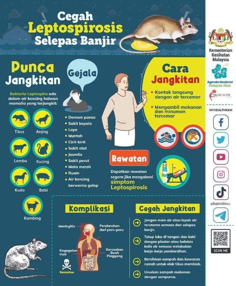
According to the guidelines given by the Ministry of Health Malaysia (2011), health education activities are conducted to create awareness of leptospirosis. Electronic media, printed materials and interpersonal such as conversations with colleagues or family are some of the strategies made by the ministry to spread awareness to the public. 

Figure 7. Infographic of leptospirosis


Then, risk assessments of possible contaminated water sources/bodies are conducted to know the safety of the water source and the availability of facilities to prevent rodents from roaming in places such as recreational parks as shown in Figure 8. Other than that, after assessing an area and the area is found to be contaminated with leptospirosis, health hazard signage is placed to warn the public as shown in Figure 9.


Figure 8. Example of risk assessment for recreational park


Figure 9. Example of hazard signage


Next, individuals who are exposed to the contaminated water or soil are advised to wear waterproof protective clothing, cover skin lesions with waterproof dressings, wash the wound with clean water immediately after exposure to leptospirosis and seek medical treatment if there is any severe symptom occurring. Then, the public is advised to keep homes free from rodents and is urged to vaccinate their pets. Next, the ministry also promotes interagency collaboration such as PERHILITAN to maintain cleanliness in relevant environmental settings, especially rodent control and promote cleanliness campaigns in recreational and housing areas.

    Leptospirosis had only been declared as a notifiable disease as of 9th December 2010 due to unknown exact disease burden in Malaysia even though the disease has existed since the 1920s. Leptospirosis has been listed as an infectious disease under Prevention and Control of Infectious Diseases Act 1988 which includes the administration of authorized officers, police and customs, prevention of importation of leptospirosis, control of the spread of leptospirosis, offence and penalties (Prevention And Control of Infectious Diseases Act 1988, 2020).


Conclusion 

In conclusion, leptospirosis is still one of the diseases with a high rate of infections, especially during climate change that causes floods. Awareness of leptospirosis should be disseminated to the public to avoid becoming infected by pathogenic or saprophytic Leptospires. Current guidelines on leptospirosis should be updated accordingly to avoid a future outbreak.

Sunday, 19 June 2022

References


Abd Wahil, M. S., Syed Nor, S. F., Chin Mun, W., Ahmad, N., Inche Zainal Abidin, N. D., Abd Ghani, S. R., Sivaratnam, L., Syed Rusli, S. A. S., Abdul Wahab, A. A., Jaafar, S. J., & Daud, F. (2021). Visitors’ Perception of the Effectiveness of Leptospirosis Health Hazard Warning Signage in Recreational Parks, Hulu Langat District, Malaysia. ASM Science Journal, 14, 1–12. https://doi.org/10.32802/asmscj.2020.653


Bharti, A. R., Nally, J. E., Ricaldi, J. N., Matthias, M. A., Diaz, M. M., Lovett, M. A., Levett, P. N., Gilman, R. H., Willig, M. R., Gotuzzo, E., Vinetz, J. M., & Peru-United States Leptospirosis Consortium (2003). Leptospirosis: a zoonotic disease of global importance. The Lancet. Infectious diseases, 3(12), 757–771. https://doi.org/10.1016/s1473-3099(03)00830-2


Benacer, D., Thong, K. L., Verasahib, K. B., Galloway, R. L., Hartskeerl, R. A., Lewis, J. W., & Mohd Zain, S. N. (2016). Human Leptospirosis in Malaysia: Reviewing the Challenges After 8 Decades (1925-2012). Asia-Pacific journal of public health, 28(4), 290–302. https://doi.org/10.1177/1010539516640350


Caimi, K., & Ruybal, P. (2020). Leptospira spp., a genus in the stage of diversity and genomic data expansion. Infection, genetics and evolution : journal of molecular epidemiology and evolutionary genetics in infectious diseases, 81, 104241.


Ellis W. A. (2015). Animal leptospirosis. Current topics in microbiology and immunology, 387, 99–137. https://doi.org/10.1007/978-3-662-45059-8_6


Fann, R. J., Vidya, R. R., Chong, H. E., Indralingam, V., & Christopher Chan, W. S. (2020). Clinical presentations and predictors of mortality for leptospirosis - A study from suburban area in Malaysia. The Medical journal of Malaysia, 75(1), 52–56.


Fletcher, W. (1928). Recent work on leptospirosis, tustsugamushi disease and tropical typhus in the Federal Malay States. Transaction of the Royal Society of Tropical Medicine and Hygiene, 21(4), 265-288. https://doi.org/10.1016/S0035-9203(28)90019-X

Garba, B., Bahaman, A. R., Khairani-Bejo, S., Zakaria, Z., & Mutalib, A. R. (2017) Retrospective study of leptospirosis in Malaysia. Ecohealth. 14(2), 389–98. https://doi.org/10.1007/s10393-017-1234-0.


Garba, B., Bahaman, A. R., Bejo, S. K., Zakaria, Z., Mutalib, A. R., & Bande, F. (2018). Major epidemiological factors associated with leptospirosis in Malaysia. Acta Tropica, 178, 242–247. https://doi.org/10.1016/j.actatropica.2017.12.010


Goris, M. G., & Hartskeerl, R. A. (2014). Leptospirosis serodiagnosis by the microscopic agglutination test. Current protocols in microbiology, 32, . https://doi.org/10.1002/9780471729259.mc12e05s32


Haake, D. A., & Levett, P. N. (2014). Leptospirosis in humans. Current Topics in Microbiology and Immunology, 65–97. https://doi.org/10.1007/978-3-662-45059-8_5 


Inada, R., Ido, Y., Hoki, R., Kaneko, R., & Ito, H. (1916). The etiology mode of infection and specific therapy of Weil’s disease. Journal of Experimental Medicine, 23(3), 377-402.


Institute for International Cooperation in Animal Biologics, Center for Food Security and Public Health. Leptospirosis (2005). Institute for International Cooperation in Animal Biologics, Center for Food Security and Public Health.


Jabatan Kesihatan WP Kuala Lumpur & Putrajaya. (2022, January 6). #TIPSBANJIR: PENYAKIT KENCING TIKUS. https://www.mkn.gov.my/web/ms/2022/01/04/tipsbanjir-penyakit-kencing-tikus/

Karpagam, K. B., & Ganesh, B. (2020). Leptospirosis: a neglected tropical zoonotic infection of public health importance-an updated review. European journal of clinical microbiology & infectious diseases : official publication of the European Society of Clinical Microbiology, 39(5), 835–846. https://doi.org/10.1007/s10096-019-03797-4

Koay, T. K., Nirmal, S., Noitie, L., & Tan, E. (2004). An epidemiological investigation of an outbreak of leptospirosis associated with swimming, Beaufort, Sabah. The Medical journal of Malaysia, 59(4), 455–459.


Leptospirosis information center. Leptospirosis Information. (2021, July 8). Retrieved June 1, 2022, from http://www.leptospirosis.org/topic.php?t=10 


Levett P. N. (2001). Leptospirosis. Clinical microbiology reviews, 14(2), 296–326. https://doi.org/10.1128/CMR.14.2.296-326.2001 


Lim, J. K., Murugaiyah, V. A., Ramli, A. S., Rahman, H. A., Mohamed, N. S. F., Shamsudin, N. N., & Tan, J. C. (2011). A case study: leptospirosis in Malaysia.

Madhu S, Aparna Y. Leptospirosis: Epidemiology, diagnosis and control. J Infect Dis Antimicrob Agents (2008); 25(4): 93-101

McCrumb, F. R., Jr, Stockard, J. L., Robinson, C. R., Turner, L. H., Levis, D. G., Maisey, C. W., Kelleher, M. F., Gleiser, C. A., & Smadel, J. E. (1957). Leptospirosis in Malaya. I. Sporadic cases among military and civilian personnel. The American journal of tropical medicine and hygiene, 6(2), 238–256.

Ministry Of Health Malaysia. (2011). Guidelines for the diagnosis, management, prevention and control of leptospirosis in Malaysia.

Prevention And Control Of Infectious Diseases Act 1988, (2020). https://lom.agc.gov.my/ilims/upload/portal/akta/outputaktap/1687632_BI/Act%20342%20-%20Draf%20Bersih.pdf

Shafei, M.N., Sulong, M.R., Yaacob, N., Hassan, H., Mohamad, W.M., Daud, A., Ismail, Z., & Abdullah, M.R. (2012). Seroprevalence of Leptospirosis among Town ServiceWorkers in Northeastern State of Malaysia. International journal of collaborative research on internal medicine and public health, 4, 395-403.

Silva MV, Camargo ED, Batista L, Vaz AJ, Brandão AP, Nakamura PM, Negrão JM. Behaviour of specific IgM, IgG and IgA class antibodies in human leptospirosis during the acute phase of the disease and during convalescence. J Trop Med Hyg 1995;98(4):268-272.

The Leptospirosis Information Center [homepage on the Internet]. Survival of leptospires in the environment. The Leptospirosis Information Center; [updated 2010 May 28; cited 2022 June 1]. Available from: http://www.leptospirosis.org/topic.php?t=10


Victoriano, A. F., Smythe, L. D., Gloriani-Barzaga, N., Cavinta, L. L., Kasai, T., Limpakarnjanarat, K., Ong, B. L., Gongal, G., Hall, J., Coulombe, C. A., Yanagihara, Y., Yoshida, S., & Adler, B. (2009). Leptospirosis in the Asia Pacific region. BMC infectious diseases, 9, 147. https://doi.org/10.1186/1471-2334-9-147


World Health Organization. (‎2003)‎. Human leptospirosis: guidance for diagnosis, surveillance and control. World Health Organization.


Zainudin A. (2015) Epidemiology and Current Situation of Leptospirosis in Malaysia. Persidangan Kesihatan Persekitaran Pihak Berkuasa Tempatan, Labuan: Ministry of Health Malaysia.

Summary

  Leptospirosis is one of the important diseases in Malaysia. The study of leptospirosis is extensive and has also been studied in various c...